


Assessment of ductus arteriosus morphology by echocardiography compared with digital subtraction angiography in children with pulmonary atresia and ventricular septal defect
- University of Health Sciences – Vietnam National University Ho Chi Minh City, Vietnam
- Children’s Hospital 1, Ho Chi Minh City, Vietnam
Abstract
Objective: To describe the morphological characteristics of the ductus arteriosus in patients with pulmonary atresia with ventricular septal defect (PA-VSD), and to evaluate the concordance between echocardiography and digital subtraction angiography (DSA), as well as the effect of prostaglandin E₁ (PGE₁) on improving oxygen saturation.
Materials and methods: Clinical data and morphological characteristics of the ductus arteriosus were collected from medical records, color Doppler echocardiography, and DSA. The evaluated characteristics included the number of ductus arteriosus, site of origin, course, diameter at the aortic end, and diameter at the pulmonary end. Categorical variables were presented as frequencies and percentages, while continuous variables were presented as mean ± standard deviation. Differences in ductal diameters between the two imaging modalities were analyzed using paired comparison tests. Given the small sample size, all analyses were interpreted as exploratory.
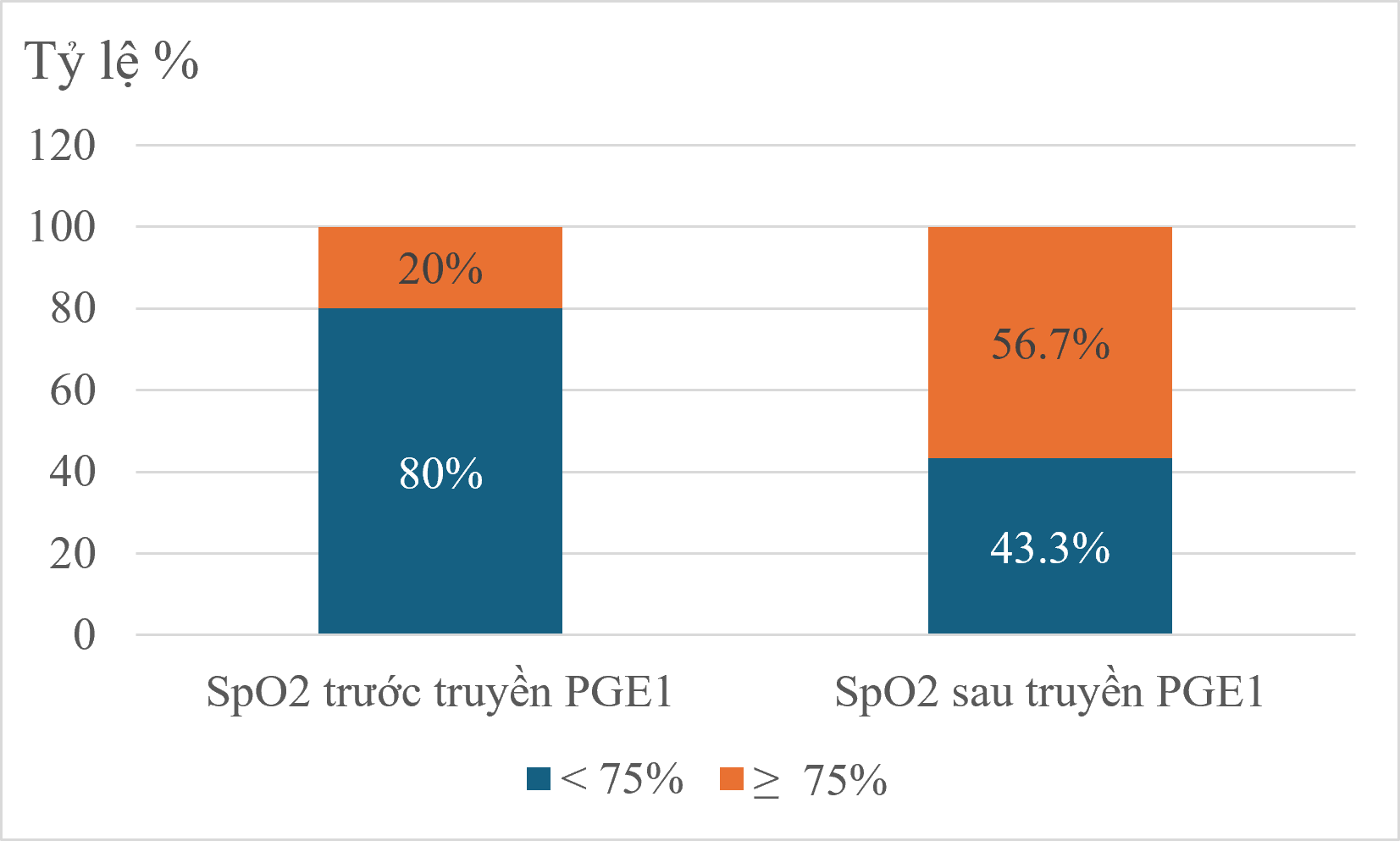
Results: All patients had a single ductus arteriosus. On echocardiography, the ductus arteriosus originated from the descending aorta in 53.3% of cases, from the brachiocephalic artery in 36.7%, and from the subclavian artery in 10.0%. On DSA, the corresponding proportions were 50.0%, 40.0%, and 10.0%, respectively. A vertical course was observed in 56.7% of cases on both imaging modalities. The aortic-end diameters measured by echocardiography and DSA were 3.12 ± 0.97 mm and 3.86 ± 1.43 mm, respectively, with no statistically significant difference in exploratory analysis. The pulmonary-end diameters were 2.18 ± 0.62 mm and 1.98 ± 0.83 mm, respectively, with no statistically significant difference. All patients were admitted due to cyanosis. Pneumonia was the most common comorbidity, accounting for 40.0% of cases. After PGE₁ infusion, the proportion of patients with SpO₂ ≥75% increased from 20.0% to 56.7%.
Conclusion: Echocardiography is a useful initial imaging modality for assessing the morphological characteristics of the ductus arteriosus in children with pulmonary atresia with ventricular septal defect, particularly regarding ductal number, course, and site of origin. DSA remains necessary in patients with complex anatomy and before intervention. Changes in SpO₂ after PGE₁ infusion should be regarded as supportive clinical information rather than a primary study outcome.