


Efficient brain tumor detection in MRI scans using YOLOv11 with pretrained backbones

- School of Computer Science and Engineering, The Saigon International University, Ho Chi Minh City 700000, Vietnam
- Department of Information Technology, FPT University, Ho Chi Minh City 700000, Vietnam
- International University - Viet Nam National University Ho Chi Minh City
Abstract
[Background] Precise object detection within medical imaging constitutes a fundamental requirement for computer-aided diagnostic systems, particularly for the localization of brain tumors. Brain tumors present significant diagnostic challenges due to their heterogeneity in shape, size, texture, and anatomical location across varying Magnetic Resonance Imaging (MRI) modalities. While manual interpretation by clinical specialists is the gold standard, it remains labor-intensive, time-consuming, and subject to inter-observer variability. Consequently, automated deep learning systems have emerged as vital tools for rapid screening. However, balancing detection accuracy with computational efficiency remains a critical challenge for real-world clinical deployment, particularly when moving beyond simple classification to precise spatial localization.
[Objective] This study investigates the performance efficacy of integrating distinct Convolutional Neural Network (CNN) backbones into the state-of-the-art YOLOv11 architecture to optimize tumor detection in MRI scans. Our primary aim is to systematically evaluate how variations in feature extraction architecture influence the critical trade-off between localization precision and computational complexity.
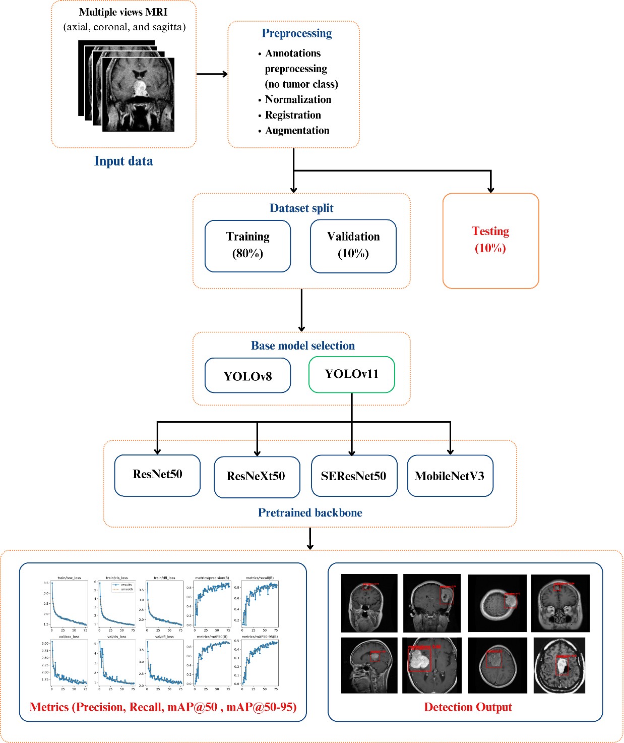
[Methods] Four model variants were engineered and tested by substituting the native YOLOv11 backbone with structurally diverse pretrained networks: ResNet50, ResNeXt50, SEResNet50 (incorporating attention mechanisms), and MobileNetV3-Large (optimized for mobile architectures). These models were trained and validated on a manually annotated dataset comprising three primary tumor classes: glioma, meningioma, and pituitary tumors. Ground truth bounding boxes were strictly delineated to capture exact tumor spatial extents. Performance was rigorously quantified utilizing standard object detection metrics—Precision, Recall, [email protected], and [email protected]:0.95—alongside an evaluation of model parameter counts to assess computational weight.
[Results] Comparative analysis reveals a distinct performance dichotomy dictated by backbone design. Deeper architectures—specifically ResNet50 and SEResNet50—yield superior feature extraction capabilities, maximizing detection accuracy and achieving the highest overall mAP scores. Conversely, MobileNetV3 demonstrates a highly favorable efficiency-performance trade-off. Despite a significantly reduced parameter count and lower computational overhead, the MobileNetV3-backed model retains competitive mAP scores, proving its capability to accurately localize abnormalities efficiently.
[Conclusion] These findings establish distinct deployment strategies tailored to clinical needs. ResNet-based backbones are optimal for precision-critical diagnostic tasks where computational resources are abundant, whereas MobileNetV3 offers a viable, lightweight alternative for implementation in resource-constrained clinical hardware or real-time preliminary screening. Future work will focus on validating these lightweight architectures across multi-center datasets to ensure broader generalizability.